|
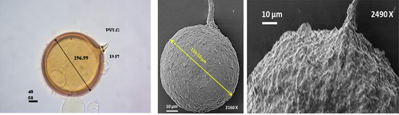
Shape: Spores are globose to sub-globose in shape and sometime slightly oval with a smooth surface.
Color: Spores are mostly hyaline; however, some of the mature spores appear light brown to pale yellow in colour. Spores show very less or no reaction with both PVLG (Polyvinyl alcohol acid glycerol) and Melzer's:PVLG.
Average diameter: 140.15 (210.76) to 320.18 μm
Spore after reaction with PVLG
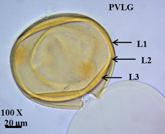
Spore wall: Three layers, L1, L2 and L3. L1 seen as a mucilaginous hyaline layer. L2 is laminated and L3 adheres to L2 and is flexible and smooth.
L3 does not show strong reaction towards Melzer's: PVLG.
Spore wall layer-1 (L1): Outermost mucilaginous hyaline layer, showing no reaction in Melzer's: PVLG. The average thickness of this layer lies between 0.5 and 1.75 μm.
Spore wall layer-2 (L2): Laminated layer next to L1. The layer is smooth and the thickest. The layer is slightly hyaline and shows no or very less reaction with Melzer's: PVLG. The average thickness of this layer lies between 1.5 and 4.5 μm.
Spore wall layer-3 (L3): This layer lies closely adhered to the innermost sub-layer of L2. L3 is flexible and smooth with an average thickness of 0.2 and 0.6μm.
L3 does not show strong reaction towards Melzer's: PVLG.
Subtending hypha: Mostly cylindrical to slightly flared hyaline subtending hyphae seen. The width of the subtending hyphae at the spore bases ranges from 2.54-3.83 μm.
The subtending hyphae consists of two wall layers hyphal wall layer 1 (HWL-1) and hyphal wall layer 2 (HWL-2), that are continuous to the L2 and L3 of the spore, respectively.
|